Related Documentation:
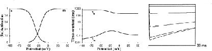
The P-type Ca2+ channel is a high-threshold, very slowly inactivating channel first described in the Purkinje cell [7]. A complete whole-cell patch clamp study of this channel in freshly dissociated rat Purkinje cells was done by [8]; this provided us with all the data necessary to model the P-type calcium (CaP) current (Fig. 2C). Initial versions of the model were run with equations based on averaged data [8] but, confirming our experience in other systems [2], we found that equations based on data from a single preparation (Figs. 5C and 6C in [8]) made Ca2+ spiking in the model more robust. These data do not support multiple activation states for the CaP channel because there does not seem to be any delay in activation [5] and the steady-state activation curve could be fitted by a Boltzmann-style curve with power 1. Therefore activation of CaP has been modeled with a single gate. This is in contrast to equations for other mammalian Ca2+ currents, which usually show some delay in activation [1, 6].
Recently, [9, 10] also reported CaP channel data on the basis of cell-attached patch clamps of guinea pig Purkinje cells. These results seem to be in accord with the data reported by [8] for activation; the threshold of activation especially is very similar (-41 mV in 2 mM Ca2+ reported by Usowicz et al. [9] vs. -45 to -40 mV in 5 mM Ba2+ [8]. However, Usowicz et al. [10] show little or no inactivation of CaP current, whereas [8] declares that there is a slow inactivation. Other authors claim that there might be several time constants of inactivation for CaP current [4] and to our knowledge a possible Ca2+-dependent inactivation, as found in other high-threshold Ca2+ channels [3] has not been completely excluded. The model used the slow inactivation suggested by [8] (Fig. 2C).
[1] C Chen and P Hess. Mechanisims of gating of t-type calcium channels. Journal of General Physiology, 96:603–630, 1990.
[2] E De Schutter, JD Angstadt, and RL Calabrese. A model of graded synaptic transmission for use in dynamic network simulations. Journal of Neurophysiology, 69:1225–1235, 1993.
[3] AP Fox, MC Nowycky, and RW Tsien. Kinetic and pharmacological properties distinguishing three types of calcium current in chick sensory neurones. Journal of Physiology (Lond.), 394:149–172, 1987.
[4] P E Hockberger and S C Nam. Purkinje cell calcium currents: Are they unique? Society for Neuroscience Abstracts, 17:1518, 1991.
[5] A Hodgkin and A Huxley. A quantitative description of membrane current and its application to conduction and excitation in nerve. Journal of Physiology (Lond.), 117:500–544, 1952.
[6] AR Kay and RKS Wong. Calcium current activation kinetics in isolated pyramidal neurons of the ca1 region of the mature guinea-pig hippocampus. Journal of Physiology (Lond.), 392:603–616, 1987.
[7] RR Llinás and M Sugimori. Electrophysiological properties of in vitro Purkinje cell somata in mammalian cerebellar slices. Journal of Physiology (Lond.), 305:171–195, 1980.
[8] LJ Regan. Voltage-dependent calcium currents in Purkinje cells from rat cerebellar vermis. Journal of Neuroscience, 11:2259–2269, 1991.
[9] MM Usowicz, M Sugimori, B Cherksey, and RR Llinás. Characterization of P-type calcium channels in cerebellar Purkinje cells. Society for Neuroscience Abstracts, 18:974, 1992.
[10] MM Usowicz, M Sugimori, B Cherksey, and RR Llinás. P-type calcium channels in the somata and dendrites of adult cerebellar Purkinje cells. Neuron, 9:1185–1199, 1992.